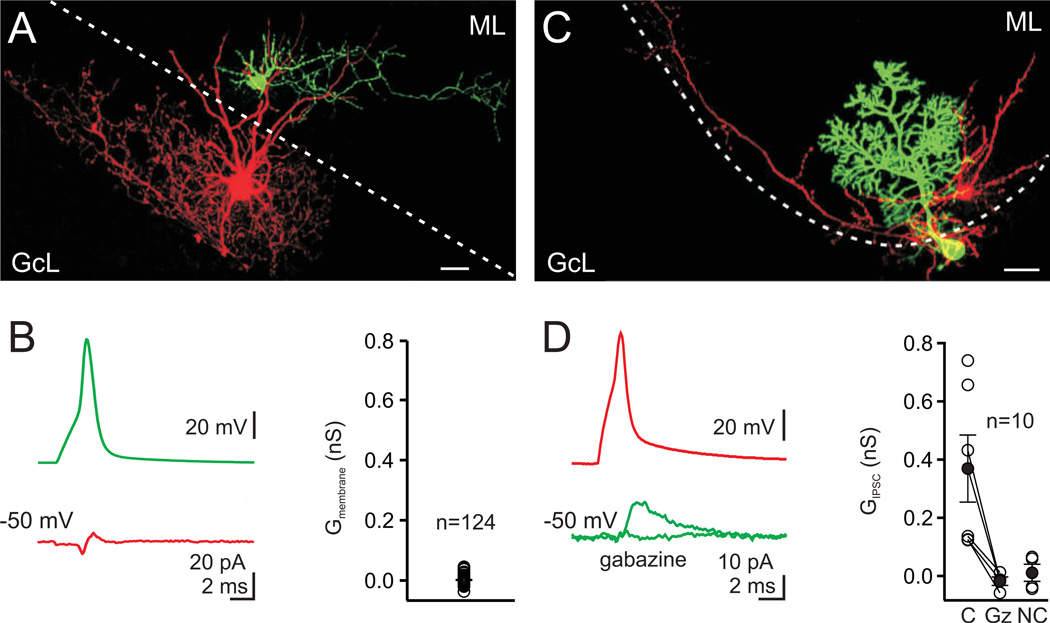
Figure 6. Paired recordings show no connections between molecular layer interneurons and Golgi cells.
A. A MLI cell filled with Alexa 488 (green) and a Golgi cell filled with Alexa 594 (red), were imaged with 2-photon microscopy. Scale bar=20 µm, ML=molecular layer, GcL=granule cell layer, dotted line is the boundary of the molecular layer. B. left, Spiking the MLI (green trace) did not produce an IPSC in the Golgi cell (red trace, average of 70 consecutive trials, inflection is a capacitative electrical artifact). right, the average membrane conductance measured from 61 unconnected pairs was 0.001 nS. C. Paired recording from a MLI (basket cell, red) and a Purkinje cell (green). D. left, Spiking the MLI produced an IPSC in the Purkinje cell that was abolished by gabazine (5 µM). right, IPSCs were observed In 6 of 10 paired recordings between MLIs and Purkinje cells. C= connected, Gz = gabazine, NC = not connected.

