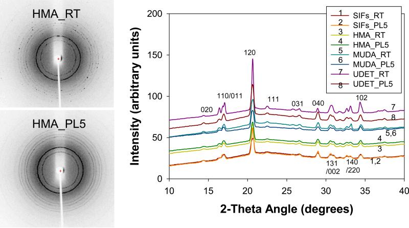
Figure 8.
Experimental 2-D (Left) and 1-D (Right) Powder X-Ray Diffraction patterns of L-alanine crystals grown from L-alanine solutions 2.7 M pH= 5.3 on glass at room temperature (RT) and using microwave heating (PL). The Miller indices corresponding to the peaks are also shown. The bell-shape in the 1-D plot is due to the background signal as also observed in the previous publications by others.

