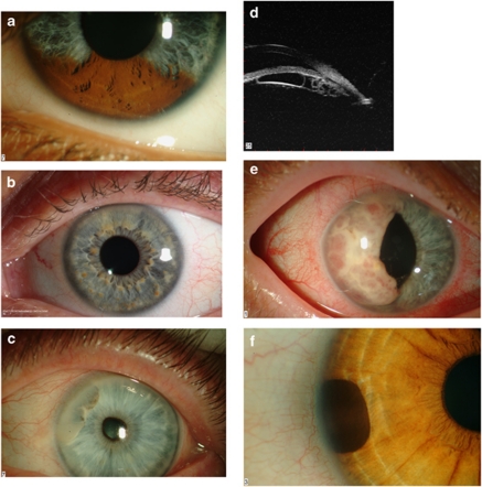
Figure 1.
(a) Sectorial pigmentation in Waardenburg syndrome. (b) Lisch nodules in neurofibromatosis type-I. Note the predominant inferior location. (c) Anterior stromal cyst containing a turbid sediment. (d) Multiple posterior pigment epithelial cysts. This patient presented with raised intraocular pressure. Note the plateaux iris configuration. (e) A large iris metastasis in a patient with an occult bronchiogenic carcinoma. (f) A peripheral iris melanocytoma.

