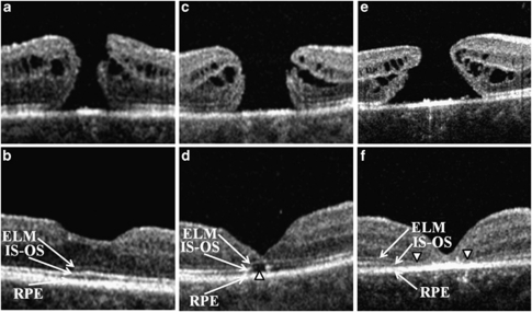
Figure 1.
Representative SD-OCT images of the three groups. Preoperative (upper images) and postoperative images at 6 weeks after the macular hole repair (lower images). SD-OCT, spectral domain optical coherence tomography; ELM, external limiting membrane; IS–OS, inner segment–outer segment junction layer; RPE, retinal pigment epithelium. First group (a, b)—ELM continuous and IS–OS continuous (ELMc/IS–OSc), second group (c, d)—ELM continuous and IS–OS discontinuous (ELMc/IS–OSd), and third group (e, f)—ELM discontinuous and IS–OS discontinuous (ELMd/IS–OSd). Arrowheads indicate disruptions in IS–OS (d) and ELM (f).

