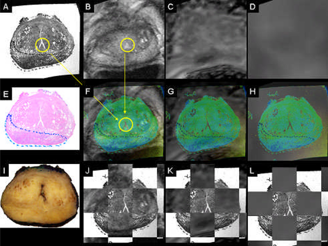
Figure 2.
Coregistered whole mount HE histology (A,E), in-vivo T2 MRI reference space (B), coregistered diffusion-weighted MRI (C), coregistered 18F-FAZA PET/CT (D), coregistered color-coded histology and respective in-vivo imaging (F-H), block face photograph (I), coregistered color-coded histology and in-vivo imaging as checkerbox (J-L). Same patient as in Figure 1.

