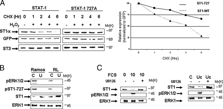
FIGURE 6.
Stability of STAT1 is modulated by the activity of ERK Kinase. A, mutant STAT1S727A degradation is significantly reduced following oxidative stress. STAT1-deficient MEF cell were transfected with wild-type STAT1 or mutant STAT1S727A or a green fluorescent protein expression vector and exposed to cycloheximide (CHX) plus H2O2 (200 μm) for the indicated times. Western blotting was carried out with the indicated antibodies. In the right panel, the results shown in A were subjected to densitometric analysis and results were normalized against green fluorescent protein (GFP) expression levels, showing wild-type STAT1α (ST1-WT, triangles) or mutant STAT1S727A (ST1-727, squares) levels. Similar results were observed in three independent experiments. B, pharmacologic inhibition of ERK activity enhances STAT1 levels. Leukemic cell lines Ramos and RL were treated with the MEK1 inhibitor U0126 (U) (1 μm) for 6 h and analyzed by Western blotting with the indicated antibodies, anti-STAT1α antibody (C-24) (ST1) or phospho-ERK (pERK). Similar results were observed in three independent experiments. Lane C, control. C, wild-type MEF cells were arrested by serum starvation for 24 h (0) and then allowed to cycle again following addition of 10% serum (10) or 10% serum plus addition of the MEK1 inhibitor U0126 (1 μm) (10 +). FCS, fetal calf serum. After 6 h cell lysates were analyzed by Western blotting with the indicated antibodies, anti-STAT1α antibody (C-24) (ST1) or phospho-ERK (pERK). Similar results were observed in three independent experiments (left panel). Wild-type MEF cells were treated with 1 × 10⁶ m urocortin (Uc) or urocortin plus the MEK1 inhibitor U0126 (1μm) (Uc +). After 6 h cell lysates were analyzed by Western blotting with the indicated antibodies, anti-STAT1α antibody (C-24) (ST1) or phospho-ERK (pERK). Similar results were observed in three independent experiments (right panel).

