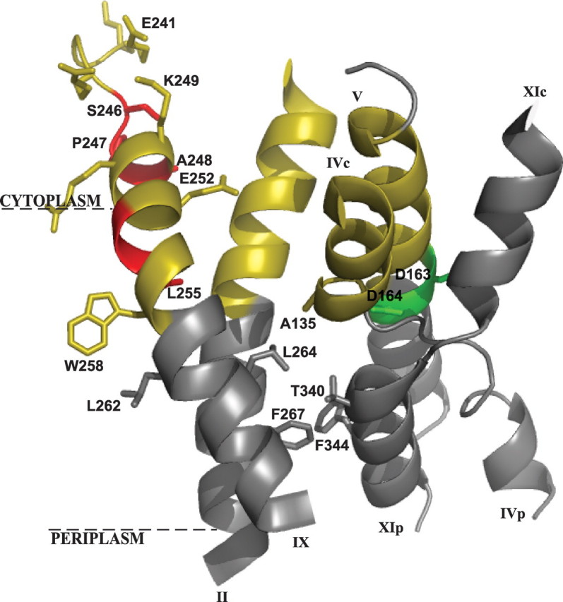
FIGURE 1.

Structure of the cytoplasmic funnel. Shown is a crystal structure-based ribbon representation of the cytoplasmic funnel (colored yellow) composed of TMSs II, IVc (c and p denote cytoplasmic and periplasmic sides, respectively), V, and IX. The TMS IV/XI assembly of short helices connected by extended chains is shown (11). The NhaA dimer interface is marked in red, the putative ion binding site is in green, and the membrane is represented by a broken line. The representation was generated using PyMOL (DeLano Scientific LLC).
