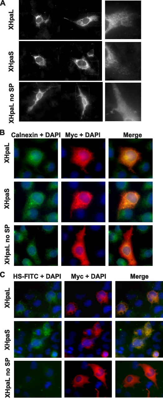
FIGURE 4.

Cellular localization of XHpaL and XHpaS. A, glioma C6 cells were transiently transfected with myc-tagged XHpaL, XHpaS, or XHpaL without signal peptide (XHpaL no SP). After 24 h, cells were fixed and stained with anti-myc antibodies. Note the reticular and pycnotic staining in XHpaL- and XHpaS-transfected cells (higher magnification on the right panel), whereas cell staining is diffuse for XHpaL no SP. B, COS-7 cells transiently transfected were stained with antibody against the myc epitope (red) and calnexin (green). 4′,6-Diamidino-2-phenylindole (DAPI) (blue) was used to detect the nucleus. C, COS-7 cells were transfected as in A. After 24 h, cells were incubated with FITC-HS for 2 h. HS is detected as green fluorescence, and heparanase expression was detected using anti-myc antibodies (red). Co-localization of heparanase and calnexin (B) and heparanase and HS (C) are detected in the merged images as yellow.
