FIGURE 5.
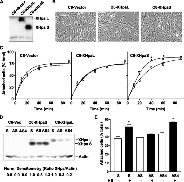
Effect of XHpaL and XHpaS on adhesion of glioma C6 cells to a HS substrate. A, Western blot of C6-stable cell lines. Cell extract obtained from C6 cells stably expressing the mock control vector (C6-vector), the long (C6-XHpaL), or the short (C6-XHpaS) myc-tagged heparanase were blotted using anti-myc antibodies. B, phase-contrast photomicrography of stable cell lines. C, cell adhesion kinetics. Cell suspensions of stable cell lines were seeded on dishes previously coated with (filled symbols) or without (open symbols) HS(50 μg/ml). At various times, the extent of adhesion was determined and expressed as percentage of the total cells seeded. Curves were fit to a one-phase exponential association (zero to top). *, p < 0.05; analysis of variance. D, stable cell lines were transfected with sense (S) or antisense oligonucleotides against exon 3 (AS) or exon 4 (AS4). After 24 h, cell extracts were analyzed by Western blot using anti-β-actin and anti-myc antibodies to detect heparanase expression. Densitometry analysis between heparanase and β-actin expression normalized against sense-transfected cells is shown at the bottom. A representative example of two independent experiments is shown. E, C6-XHpaS cells were transfected with sense and antisense oligonucleotides as explained for B. After 48 h, cell suspensions were obtained and the extent of adhesion was determined following 20 min incubation. For C and E, one each of three independent experiments are shown (*, p < 0.05; analysis of variance).

