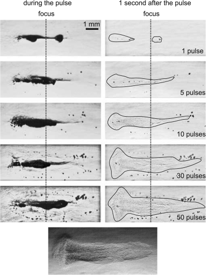
Figure 5.
High-speed camera images of lesion evolution in a polyacrylamide gel during pulsed HIFU exposures with 10 ms pulses and a 0.01 duty factor (same parameters as in Fig. 2). Images were recorded at 9.5 ms of each 10 ms pulse (left column) and 1 s after each pulse, immediately prior to the subsequent pulse (right column). A large boiling bubble forms during each 10 ms pulse (as shown in Fig. 2), but dissolves in the one second interval between pulses. Several smaller (tens of microns) residual bubbles remain between pulses and are evident in the frames on the right. These bubbles are largely pushed beyond the focus and away from the acoustic axis by the start of the subsequent pulse, collecting at the periphery of the lesion. The residual void of eroded gel is outlined in the right column for better visibility. Explosive growth of the bubble itself and also generation of high negative pressures caused by reflection of the shock wave from the bubble can contribute to tearing of the gel and lesion growth. The images were enhanced by zooming and altering the lighting to enhance the appearance of the residual void (bottom image), which contained gel pieces and infiltrated liquid and was no longer intact gel.

