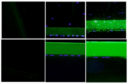
FIGURE 15.
Immunoreactivity of Descemet’s membrane (upper panel) and anterior lens capsule (lower panel) to anti-collagen type IV in negative control (left panel) and normal (middle panel) and 2-year-old (right panel) buphthalmic animal. Note the increased staining intensity in Descemet’s membrane and the anterior lens capsule in the buphthalmic animal compared to normal control (original magnification ×20, Alexa Fluor; DAPI used as nuclear stain).

