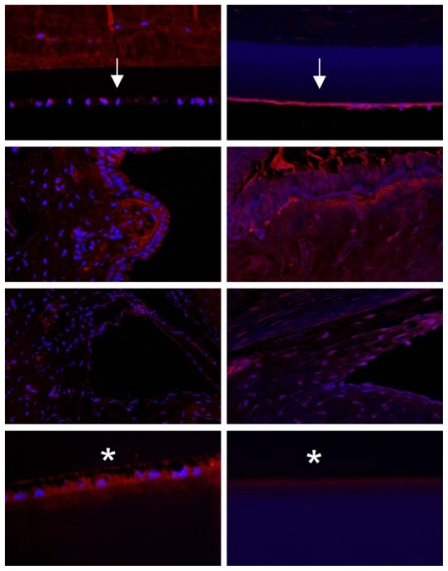
FIGURE 21.
IRBP immunofluorescence in the 5-year-old normal (left panel) and buphthalmic (right panel) rabbit in cornea (top row), ciliary epithelium (second row), angle (third row), and lens epithelium (bottom row). Note the reduced staining in most buphthalmic tissues and its absence in Descemet’s membrane (arrows) and lens capsule (*). There is slight increase in staining of corneal endothelium (original magnification ×20, Texas Red; DAPI nuclear stain).

