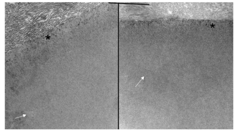
FIGURE 8.
Electron micrograph of the interface between corneal stroma in Descemet’s membrane in the normal control (left) and the buphthalmic (right) rabbit. Note the normal anterior banded zone (*) in both rabbits. The posterior nonbanded zone (arrows) in both animals appeared unremarkable (original magnification ×10,000).

