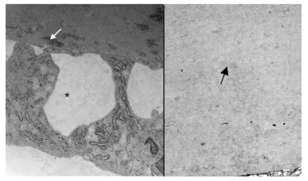
FIGURE 9.
Electron micrograph showing the posterior Descemet’s membrane and endothelium in the normal rabbit (left) and posterior part of thickened Descemet’s membrane in a 2-year-old buphthalmic rabbit (right). Left, Note the presence of abundant long-spacing collagen (white arrow) in the posterior nonbanded zone of Descemet’s membrane. The corneal endothelial cells show prominent rough endoplasmic reticulum and widened intercellular spaces (*). Right, Note the fibrillar collagenous basement membrane in the posterior nonbanded zone (black arrow) in the buphthalmic rabbit with absence of long-spacing collagen (original magnification × 12,000, and ×10,000, respectively).

