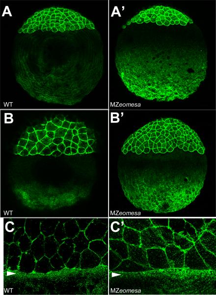
Figure 6. The actin cytoskeleton is normal is MZeomesa embryos.
Confocal projections of lateral views of phalloidin stained embryos. (A-C) wild type and (A’-C’) MZeomesa embryos. (A, A’) sphere stage (B, B’) dome stage, (C, C’) close up of marginal region at 75% epiboly, arrowheads indicate actin band in the YSL.

