
FIGURE 1.
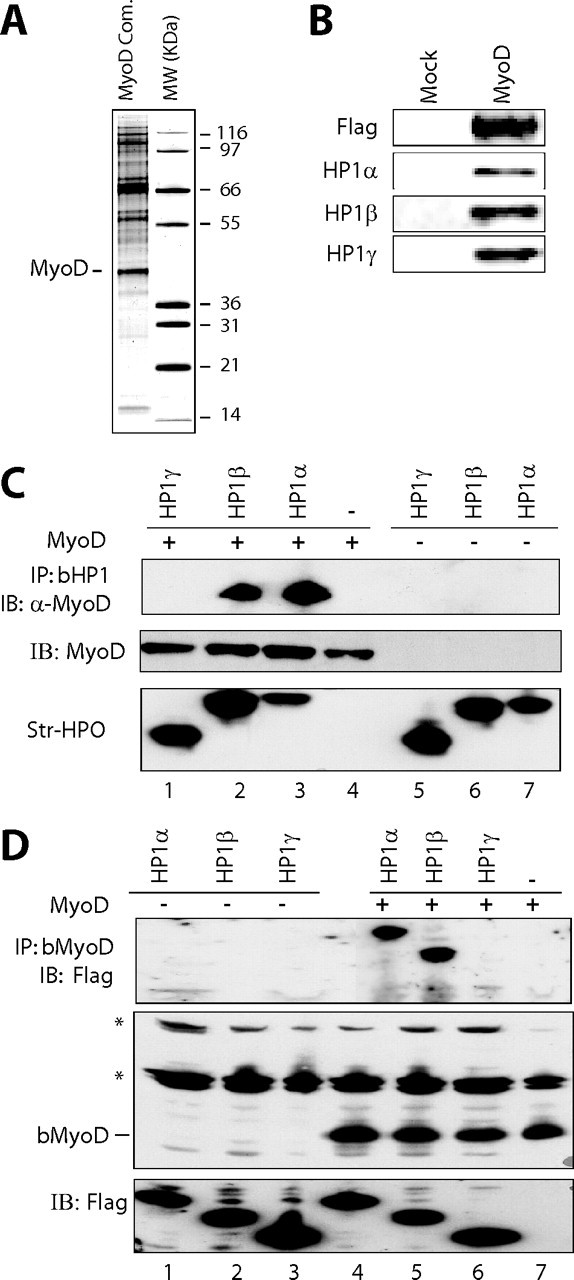
Ectopically expressed MyoD interacts with endogenous HP1 proteins. A, silver staining the double affinity-purified MyoD complex isolated from chromatin fractions in a HeLa cell line stably expressing FLAG-HA tagged MyoD. MW, protein molecular weight marker. The molecular masses of the markers are indicated. B, Western blot analysis of double purified FLAG-HA-MyoD (MyoD) or eluate from HeLa control cells (Mock). C, top panel, Western blotting with anti-MyoD on streptavidin-bead precipitates from HEK 293 cell extracts transfected with a MyoD expression vector (lanes 1–4) or with an empty vector (lanes 5–7), along with expression vectors for the biotinylated forms of HP1α (bHP1α)(lanes 3 and 7), HP1β (lanes 2 and 6), HP1γ (lanes 1 and 5), or the empty vector (lane 4). Middle and bottom panels, quantity control of MyoD detected with an anti-MyoD antibody (middle panel) and biotinylated HP1 detected using streptavidin-horseradish peroxidase conjugate (bottom panel) in the inputs. D, top panel, Western blotting of anti-FLAG-HP1 of streptavidin bead precipitates from HEK 293 cell extracts transfected with the expression vector for FLAG-HP1α (lanes 1 and 4), FLAG-HP1β (lanes 2 and 5), HP1γ (lanes 3 and 6), or with the empty vector (lane 7), along with expression vectors for the biotinylated form of MyoD (lanes 4–7) or the empty vector (lanes 1–3). Middle and bottom panels, quantity control of biotinylated MyoD (bMyoD) detected using streptavidin-horseradish peroxidase conjugate (middle panel) and that of FLAG-HP1 protein in the inputs as detected with an anti-FLAG antibody (bottom panel). *, endogenous biotinylated proteins. IB, immunoblot.