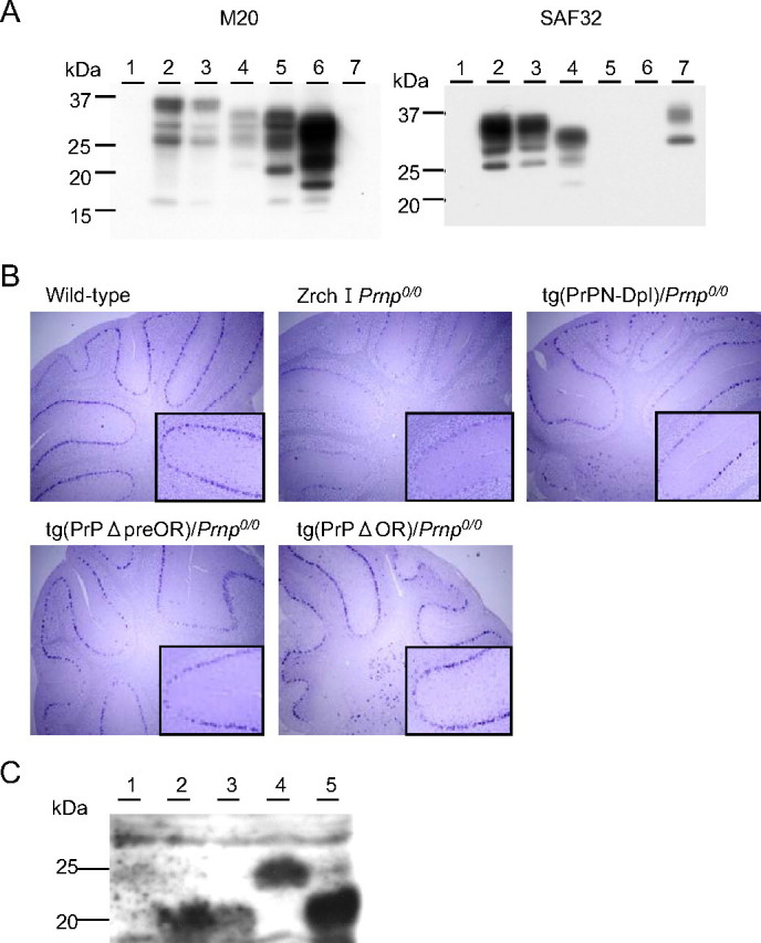
FIGURE 2.

A, Western blotting of the cerebella of tg(PrPN-Dpl)/Prnp0/0, tg(PrPΔpreOR)/Prnp0/0, and tg(PrPΔOR)/Prnp0/0 mice. 30 μg of the total proteins were loaded onto each lane. Lane 1, Zrch I Prnp0/0 mice; lane 2, wild-type mice; lane 3, Zrch I Prnp0/+ mice; lane 4, tg(PrPΔpreOR)/Prnp0/0 mice; lane 5, tg(PrPΔOR)/Prnp0/0 mice; lane 6, tg(MHM2Δ23-88)/Prnp0/0 mice; lane 7, tg(PrPN-Dpl)/Prnp0/0 mice. B, in situ hybridization of the cerebella of wild-type, Zrch I Prnp0/0, tg(PrPN-Dpl)/Prnp0/0, tg(PrPΔpreOR)/Prnp0/0, and tg(PrPΔOR)/Prnp0/0 mice. Purkinje cells in Zrch I Prnp0/0 mice show background staining with the PrP cRNA probe. In contrast, strongly stained Purkinje cells are observed in wild-type, tg(PrPN-Dpl)/Prnp0/0, tg(PrPΔpreOR)/Prnp0/0, and tg(PrPΔOR)/Prnp0/0 mice. Magnification, ×10; inset magnification, ×50. C, Western blotting of the PNGase F-treated homogenates of the cerebella from wild-type (lane 1, 100 μg of the total proteins), Ngsk Prnp0/0 (lane 2, 100 μg), Ngsk Prnp0/+ (lane 3, 100 μg), tg(PrPN-Dpl)/Prnp0/0 (lane 4, 200 μg), and tg(Dpl32)/Prnp0/0 mice (lane 5, 100 μg) using anti-Dpl FL176 antibodies.
