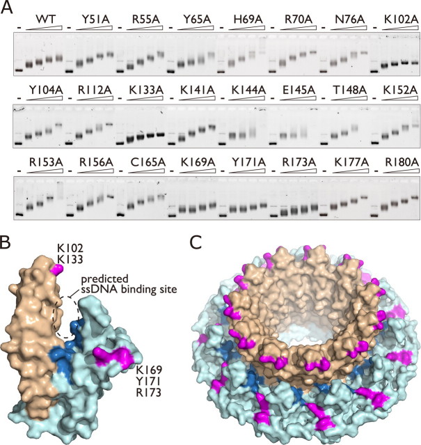
FIGURE 1.
Alanine scan mutagenesis of Rad521-212. A, EMSA. Alanine point mutants (0, 0.5, 1, 1.5, or 2 μm) of Rad521-212 were incubated with 15 μm negatively supercoiled plasmid DNA (pGsat4, 3.2 kilobases), and complexes were fractionated through an agarose gel. WT, wild type. B, surface view of the Rad521-212 protomer. The stem and domed cap regions are colored light brown and light blue, respectively. Lys-102 and Lys-133 (magenta) are located at the rim of the stem region, whereas Lys-169, Tyr-171, and Arg-173 (also magenta) are located at the edge of the domed cap region. The previously identified ssDNA binding residues (Arg-55, Tyr-65, Lys-152, Arg-153, Arg-156, dark blue) are clustered at the bottom of the groove formed between the stem and domed cap regions. C, surface view of the Rad521-212 undecameric ring. All structural figures were created using the PyMOL program.

