
FIGURE 5.
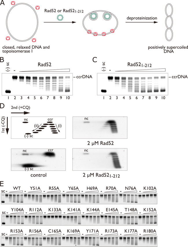
Positive supercoiling activity of Rad52 and Rad521-212. A, schematic diagram of the topoisomerase I-mediated relaxation assay. B and C, one-dimensional gel electrophoresis of DNA topoisomers generated in the presence of Rad52 (B) and Rad521-212 (C). In these reactions the protein concentrations were increased from 0 to 4 μm in 0.5 μm increments. ccrDNA denotes covalently closed, relaxed DNA. D, two-dimensional gel electrophoresis of DNA topoisomers generated in the presence of Rad52 and Rad521-212. The 2 μm Rad52 reaction (B, lane 6), resolved in a two-dimensional gel, is shown in D, top right. Similarly, the 2 μm Rad521-212 reaction (C, lane 6), resolved in a two-dimensional gel, is shown in D, bottom right. The top left figure in D is a schematic diagram of the relative positions of covalently closed, relaxed DNA (ccr), nicked circular DNA (nc), positively supercoiled topoisomers (i), and negatively supercoiled topoisomers (ii) resolved by two-dimensional gel electrophoresis. E, alanine scan mutagenesis of Rad521-212. The mutant proteins (0, 0.5, 1, 1.5, or 2 μm) were incubated with 30 μm of covalently closed, relaxed plasmid DNA. CQ, chloroquine.