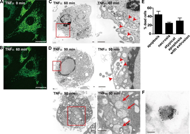
FIGURE 1.
Fragmented mitochondria are engulfed by the cytoplasmic vacuoles and extruded from c-Flip-/- MEFs during cell death. A and B, c-Flip-/- MEFs stably expressing GFP-COX IV were unstimulated (A) or stimulated with TNFα for 60 min (B). Scale bars, 25 μm. C and D, c-Flip-/- MEFs were stimulated with TNFα for 60 (C) or 90 (D) min and analyzed by transmission electron microscopy. The enlarged images of the boxed areas are shown in the right panels. The arrowheads and arrows indicate extruded and fragmented mitochondria, respectively. Scale bars, 500 nm. E, the percentages of cells showing typical apoptosis, necrosis, and atypical apoptosis characterized by numerous cytoplasmic vacuoles and mitochondrial extrusion were calculated by counting randomly selected areas (total 100-200 cells/sample). Three independently prepared samples were counted and are presented as the means ± S.D. F, c-Flip-/- MEFs were stimulated with TNFα for 90 min and then analyzed by immunoelectron microscopy using anti-COX IV antibody, followed by 10-nm colloidal gold-conjugated secondary antibody. Scale bar, 200 nm.

