FIGURE 6.
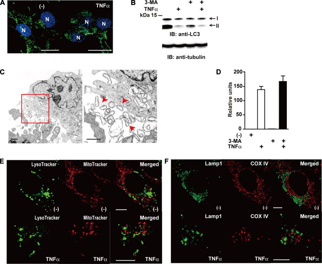
Autophagy does not play a major role in mitochondrial extrusion. A, c-Flip-/- MEFs were unstimulated or stimulated with TNFα for 90 min. Then the cells were fixed and immunostained with anti-LC3 antibody (green), and the nuclei were stained with Hoechst 33258 (blue). N, nucleus. Scale bar, 10 μm. B, c-Flip-/- MEFs were untreated or treated with TNFα, 3-MA, or TNFα plus 3-MA for 90 min. The cell lysates were analyzed by immunoblotting (IB) with anti-LC3 antibody. The arrows indicate LC3-I and LC3-II. The equal loading of the samples was verified by Western blotting with anti-tubulin antibody. The molecular mass markers are shown on the left. C, c-Flip-/- MEFs were stimulated with TNFα plus 3-MA for 90 min and analyzed by transmission electron microscopy. The enlarged image of the red box is presented in the right panel. The red arrowheads indicate extruded mitochondria. Scale bar, 1 μm. D, c-Flip-/- MEFs were untreated or treated as in B, and caspase 3 activities were measured by using fluorogenic substrates. The results are presented as the means ± S.D. of triplicate samples. E, c-Flip-/- MEFs were stimulated as in A. Then the cells were stained with LysoTracker (green) and MitoTracker (red). Scale bars, 10 μm. F, c-Flip-/- MEFs were stimulated as in A. Then the cells were fixed and immunostained with anti-Lamp1 (green) and anti-COX IV (red) antibodies. Scale bars, 10 μm.

