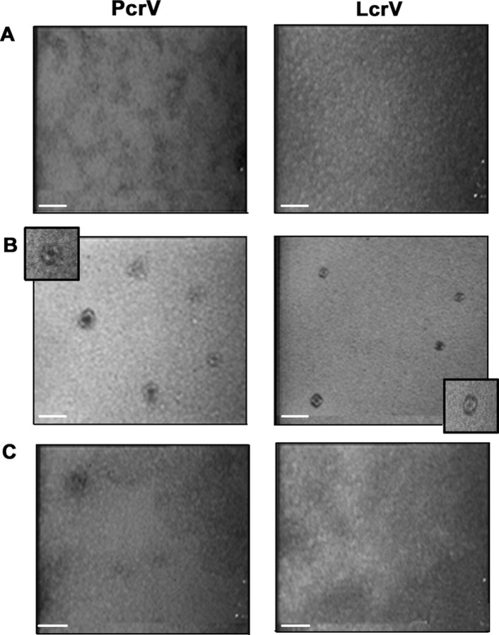
FIGURE 3.
Oligomeric V species exhibit doughnut-like shapes. Transmission electronic microscopy photographs of fractions obtained after size exclusion chromatography of untreated (A), oligomeric (B), and monomeric (C) PcrV or LcrV. All samples were coated on carbon/formar grids at a protein concentration of 0.1 mg/ml and stained with 2% uranyl acetate. No distinct structures could be observed in peaks corresponding to monomeric forms (A and C), whereas doughnut-like structures are readily observed in samples corresponding to oligomeric PcrV or LcrV (B). Scale bar corresponds to 20 nm.

