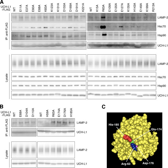
FIGURE 3.
Alanine-scanning mutagenesis of UCH-L1. A and B, lysates of COS-7 cells transfected with the indicated constructs were immunoprecipitated (IP) with anti-FLAG antibody and analyzed by immunoblotting. C, a structural model for human UCH-L1 is shown. Arg63, Glu174, Asp176, and His185 are shown in blue, green, magenta, and red, respectively, using Cn3D software (version 4.1) and NCBI structural model mmdbId:38174 (35).

