FIGURE 5.
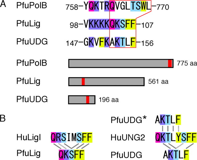
Putative PCNA-binding motif in PfuUDG. A, the PCNA-binding motifs found in PfuPolB (36) and PfuLig (35) are shown. Based on the PfuLigPCNA interaction, a PCNA-binding motif 152AKTLF156 was predicted in PfuUDG. The basic residue and the glutamine (Q), serine/threonine (S/T) and phenylalanine (F) residues are highlighted in blue, magenta, light blue, and yellow, respectively. The locations of the PCNA-binding motifs in PfuPolB, PfuLig, and PfuUDG are shown in red boxes. B, comparison of the amino acid sequences of the PCNA-binding motifs among the Ligs and UDGs from eukaryotes and Archaea. Identical and similar amino acid residues are connected by solid and broken lines, respectively.

