
FIGURE 8.
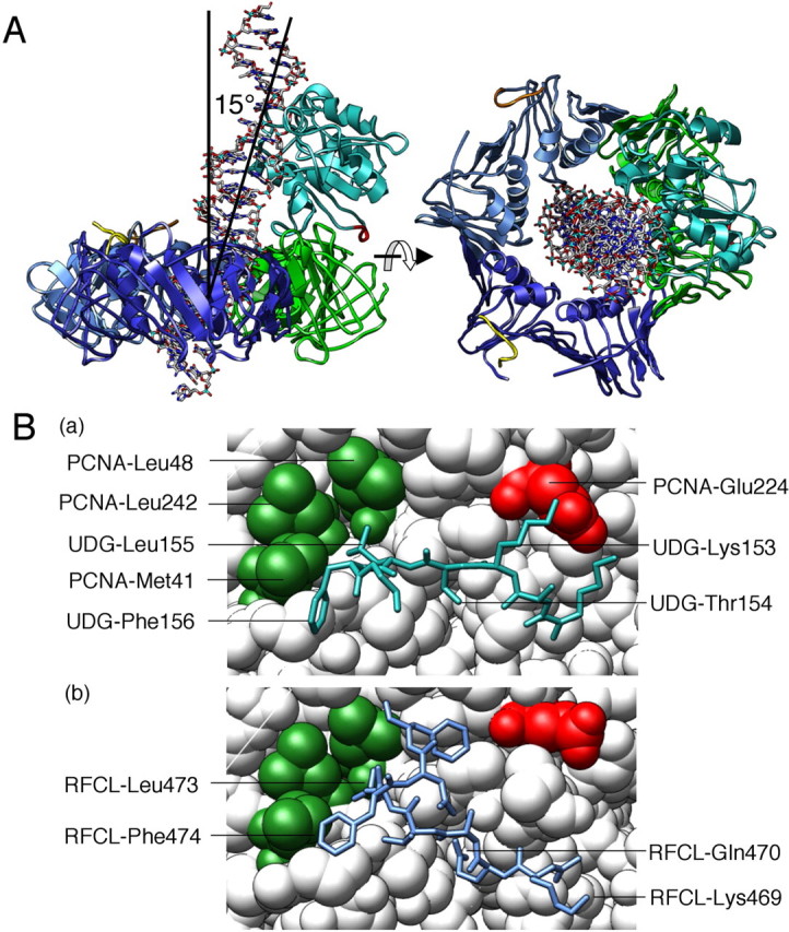
Molecular model of the PfuUDG·PCNA·DNA complex. A, the assembled model revealed that the dsDNA (stick model) complexed with PfuUDG (colored mint) pierces the hole of the PfuPCNA trimer (subunits are colored blue, dark blue, and green, respectively) when the newly identified PCNA-binding site (colored red) was properly placed at the PIP-binding site of PfuPCNA. B, close-up view of the PfuUDG PIP-box peptide (blue in the stick model) bound to PfuPCNA (a). The hydrophobic Leu-155 and Phe-156 of PfuUDG are placed in the conserved hydrophobic pocket of Leu-48 and Leu-242 in PfuPCNA. This interaction mode is clearly conserved in the interaction of PfuPCNA-RFCL as shown in (b) drawn from our previous study (43).