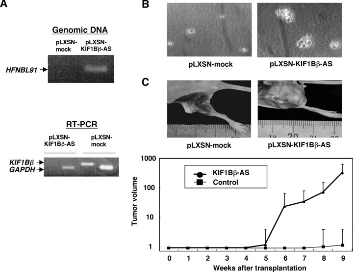
FIGURE 3.
Tumor formation in vivo. A, NMuMG cells were infected with an empty retrovirus vector (pLXSN) or with pLXSN bearing mouse antisense KIF1Bβ (pLXSN-KIF1Bβ-AS). Genomic integration of the antisense KIF1Bβ was examined by PCR (upper panel). Lower panel shows the expression levels of KIF1Bβ as examined by RT-PCR. Arrows indicate the positions of PCR products corresponding to KIF1Bβ and GAPDH. B, NMuMG cells (5 × 10⁴ cells) infected with pLXSN or pLXSN-KIF1Bβ-AS were suspended in 3 ml of 0.4% low melting agarose dissolved in culture medium, plated onto agarose bed consisting of 0.8% low-melting agarose, and incubated at 37 °C for 5 weeks. C, tumor formation in nude mice. NMuMG cells (1 × 10⁶ cells) infected with the indicated retroviruses were injected subcutaneously and tumor volumes were estimated weekly (lower panel). Upper panels show tumors generated in nude mice.

