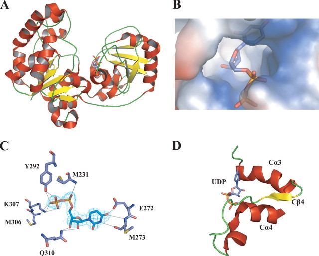
FIGURE 3.
Substrate binding in GumK. A, ribbon representation of GumK, showing UDP bound on the C-terminal face of the catalytic cleft.B, surface representation of the UDP-binding pocket. C, final (2Fo - Fc) electron density map for UDP (contoured at 1σ). Residues contacting UDP are shown as stick representations. Hydrogen bonds are depicted as dashed lines. D, GumK C-terminal α/β/α motif involved in donor substrate binding.

