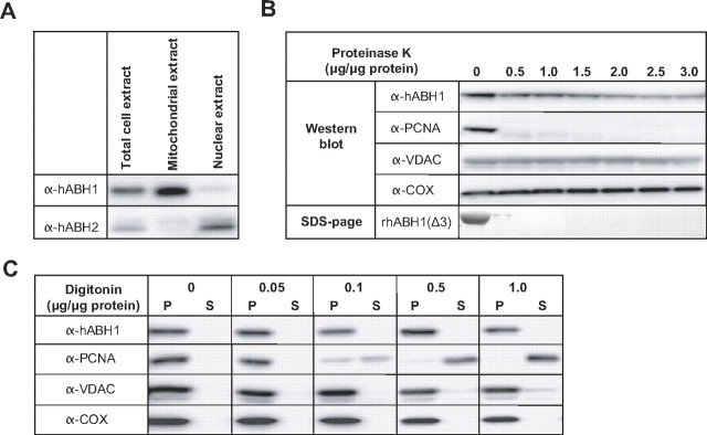
FIGURE 3.
Western blot analyses of subcellular fractions. The same amount of protein extracts (typically 20 μg) from different fractions as indicated were subjected to Western blot analysis using monoclonal antibodies against hABH1, hABH2, and PCNA, and polyclonal antibodies against VDAC (outer mitochondrial membrane marker) and COX (mitochondrial matrix marker) as indicated. A, subcellular fractionation and identification of hABH1 and hABH2 in mitochondria and nuclei, respectively. B, freshly prepared mitochondrial fractions were treated with various amounts of Proteinase K, centrifuged to remove degraded proteins, and the pellet was subjected to Western blot analysis. As a control recombinant hABH1(Δ3) was subjected to the same treatment and analyzed by SDS-PAGE. C, the mitochondria were treated with digitonin, and centrifuged to separate extracted components. The same amounts of supernatant (S) and pellet (P) were analyzed by Western blotting.

