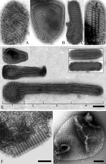
FIGURE 7.
Diverse morphological patterns of PBsomes-thylakoid membranes. A and B, flat patches; C and D, tubular shapes and vesicle topography. Interestingly, besides individual tubular fragments (E, inset), a successive process of thylakoid growth is observable. A small fragment protrudes from a base patch, which is generally bound by less organized PBsomes (E, part 1), and then develop to tubular membrane fragments with variable lengths and copies (E, parts 2–4). In addition, ordered and disorder arrangements can occasionally be imaged on the same membrane patch (F and G).

