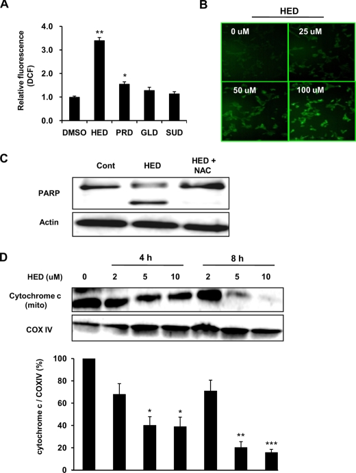
FIGURE 6.
ROS generation and cytochrome c release during HED-induced apoptosis. A, comparison of ROS generation inducted by DHA- and AA-derived dopamine adducts. The SH-SY5Y cells were treated with 10 μm dopamine adducts for 30 min and exposed to H2DCF-DA for 30 min. The fluorescence of DCF was measured by flow cytometer. B, dose-dependent ROS generation induced by HED. DCF fluorescence imaging was determined by fluorescence microscope. C, effect of antioxidant N-acetyl-l-cysteine on HED-induced PARP cleavage and accumulation of active caspase-3. 50 mm N-acetyl-l-cysteine was administrated in SH-SY5Y cells for 30 min before HED treatment. D, cytochrome c release induced by HED. The SH-SY5Y cells were treated with different concentrations of HED for 0, 4, and 8 h. The expressions of cytochrome c and cytochrome c oxidase IV (COX IV) in the mitochondrial fraction of HED-treated cells were assessed by Western blot. All of the data are shown as the means ± S.D. (n = 3) (significantly different from control: * indicates p < 0.05, ** indicates p < 0.01, and *** indicates p < 0.001. DMSO, dimethyl sulfoxide; Cont, control.

