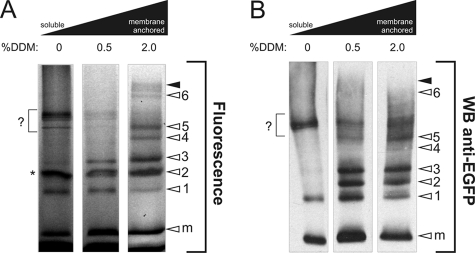
FIGURE 5.
Topology and membrane anchorage of subcomplexes in induced NDUFS3 cells. A, BN-PAGE fluorograms of membrane-solubilized proteins. Left lane represents the soluble (matrix-containing) fraction. Proteins were solubilized by treatment with DDM (concentrations are given in % (w/v)). The height of holo-CI is marked with closed arrowheads. Open arrowheads indicate NDUFS3 assembly intermediates (1–6) or monomeric subunit (m). The asterisk indicates an a-specific signal, also present in wild-type cells after contrast optimization (not shown). B, duplicate experiment as depicted in A was used for Western blotting and immunodetection with antibodies against EGFP. In this figure, the signal marked by a question mark most likely reflects sonication-induced breakdown of the peripheral arm of holo CI.

