FIGURE 5.
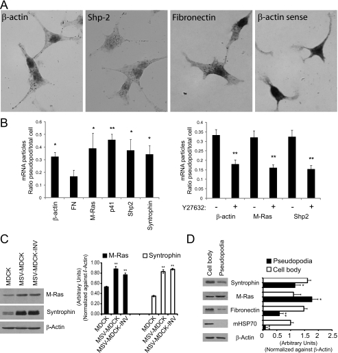
Validation and expression of pseudopodial enriched mRNAs and gene products. A, MSV-MDCK-INV cells were in situ hybridized with oligonucleotides to the antisense strands of β-actin, Shp2, and fibronectin and the sense strand of β-actin (β-actin sense). In the bright field images, dark spots indicate the presence of DIG tailed oligonucleotide bound to its complementary mRNA molecule and were absent in cells hybridized with β-actin sense oligonucleotides. B, MSV-MDCK-INV cells were in situ hybridized with oligonucleotides to β-actin, fibronectin, M-Ras, p41 protein of the Arp2/3 complex, Shp2, and α-syntrophin (left panel). Alternatively, MSV-MDCK-INV cells, untreated or treated with 20 μm Y27632 for 90 min were in situ hybridized with oligonucleotides to β-actin, M-Ras, and Shp-2 (right panel). The total number of DIG-labeled mRNA profiles or spots in pseudopodia and cell body were counted, and the average ratio of DIG-labeled mRNA profiles for pseudopodia relative to cell body is presented in graphic form for each of the indicated probes (means ± S.E.; n = 3; *, p < 0.05; **, p < 0.005 relative to FN (left) or untreated cells (right)). C, total cell lysates of MDCK, MSV-MDCK, and MSV-MDCK-INV cells were probed by Western blot for M-Ras, syntrophin, and β-actin. D, lysates from cell bodies and pseudopodia of MSV-MDCK-INV cells were probed for fibronectin, M-Ras, syntrophin, mitochondrial HSP70 (mHSP70), and β-actin. The bands were quantified by densitometry relative to β-actin. (n = 3; ±S.E.; *, p < 0.01; **, p < 0.001).

