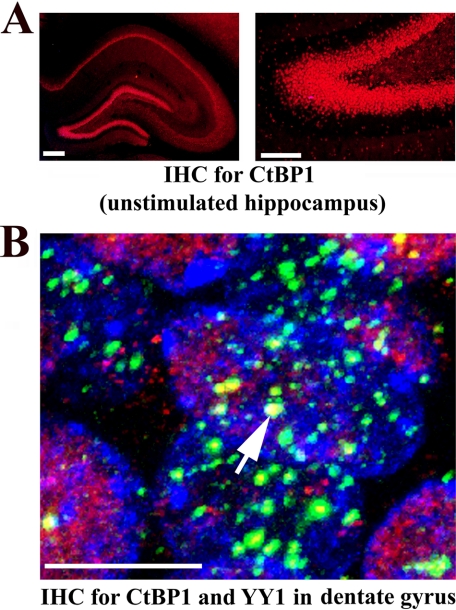
FIGURE 7.
CtBP1 is strongly expressed in the cell nuclei of the rat hippocampus and co-localizes with YY1. A, CtBP1 (in red) is expressed in the nuclei located in neuronal cell body layers of the unstimulated hippocampus. Fluorescent microscope photomicrograph on the left demonstrates CtBP1 immunolocalization throughout all areas of the rat hippocampus. Higher magnification of the same micrograph showing CtBP1 expression in a fragment of the dentate gyrus is shown on the right. Scale bars, for left photomicrograph 200 μm and for right photomicrograph 100 μm. B, CtBP1 (in red) is co-localized with YY1 expression bodies (in green) in the nuclei of the dentate gyral cells. High power confocal image of the cell nuclei from the granular layer of the unstimulated dentate gyrus is provided. One of the biggest co-localization centers is indicated with the white arrow. Cell nuclei are stained with TO-PRO-3 (in blue). Scale bar, 5 μm.

