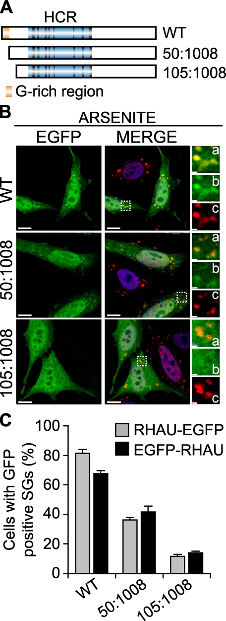
FIGURE 3.
The first N-terminal 105 amino acids of RHAU are necessary for RHAU localization in SG. A, scheme of wild-type RHAU and its deletion mutants. B, intracellular localization of RHAU N-terminal deletion mutants. HeLa cells were transfected with plasmids expressing EGFP-fused RHAU (WT) or N-terminal deletion mutants of RHAU, 50-1008 and 105-1008. After 48 h, cells were treated with 0.5 mm arsenite for 45 min, fixed, and stained for TIA-1 (red). Nuclei were visualized with DAPI (blue). Small panels show enlargements of boxed regions: merge (a), EGFP-fused fragments of RHAU (b), and TIA-1 (c). C, quantitative immunofluorescence analysis showing the percentage of transfected cells in which EGFP signals were detected in SGs. Values ± S.E.M. (standard errors of means) were derived from three independent experiments. Bar, 10 μm (1 μmin enlargements a-c). GFP, green fluorescent protein.

