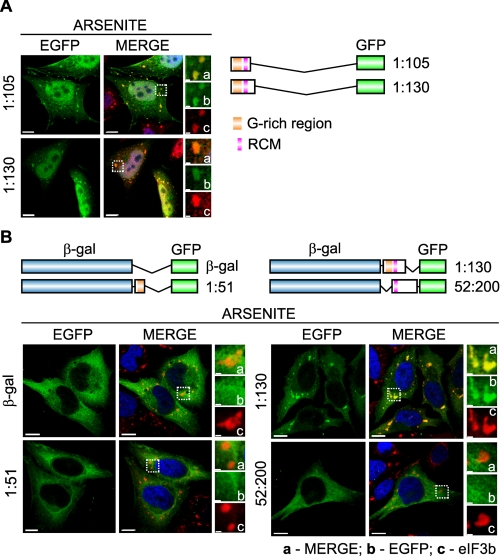
FIGURE 7.
The N-terminal RNA-binding domain is also essential and sufficient for SG localization of RHAU. A, immunofluorescent images of HeLa cells transfected with vectors expressing EGFP-tagged N-terminal fragments of RHAU, (1-105) and (1-130). After 48 h of transfection, cells were treated with arsenite (0.5 mm, 45 min) to induce SGs. The merge shows the co-localization of EGFP-tagged RHAU fragments (green) and TIA-1 (red). DAPI (blue) stains nuclei. B, immunofluorescent images of HeLa cells expressing EGFP-β-galactosidase double tagged RHAU N-terminal fragments. Cells were treated as above to induce SGs. The co-localization of EGFP (green) and eIF3b (red) is shown in the merge together with DAPI (blue). Note that only the (1-130) fragment containing an intact RNA-binding domain recruits EGFP-β-galactosidase to SGs. Enlargements of boxed regions are shown in small panels indicating merge (a), EGFP-fused RHAU N-terminal fragment (b), and eIF3b (c). Bar, 10 μm (1 μmin enlargements). GFP, green fluorescent protein.

