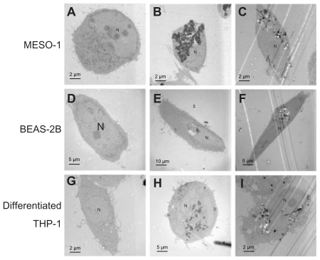
Figure 2.
TEM image of cells exposed to VGCF. Cells were exposed to varying concentrations of VGCF for 24 hours. (A, D and G) DM only. (B and H) 10 μg/mL SB. (C and I) 10 μg/mL VGCF. (E) 1 μg/mL SB. (F) 1 μg/mL VGCF.
Abbreviations: TEM, transmission electron microscope; VGCF, vapor-grown carbon fiber; SB, Sumi black; DM, dispersion medium.

