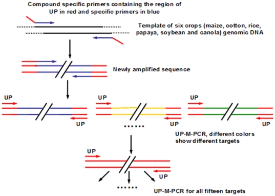
Figure 7. Amplification routine of UP-M-PCR.
Each compound specific primer contained a universal sequence at the 5′-end (red) and the specific primer at the 3′-end (blue). The amplified fragments with the primer pairs of different targets are individually marked in different colors. The amplified fragments only by the universal primer are marked in red.

