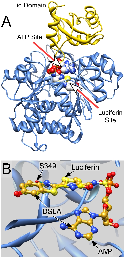
Figure 1. The structure of Japanese luciferase.
A. The backbone structure of the highest resolution structure (2D1S.pdb) is shown in ribbon format. The lid domain is shown in gold. The two ligand–binding sites are indicated with arrows and are shown occupied by the concatenated active state analog DSLA. B. The benzothiazole ring of oxyluciferin and DSLA are isosteric. The structures of Japanese luciferase with oxyluciferin +AMP bound (2D1R.pdb) and with the concatenated active state analog DSLA bound (2D1S.pdb) were superimposed. The backbone structures overlay exactly in this region and only that for 2D1R is shown. The ligands' atoms are color coded normally except carbons, which are goldenrod for DSLA and yellow for oxyluciferin and AMP.

