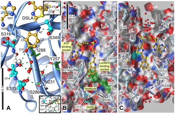
Figure 7. The structure of Japanese luciferase in the vicinity of the main photolabeled residues.
The structure is shown as a cross section through the luciferin pocket with DSLA bound in the luciferin and ATP sites (2D1S.pdb). Panel A shows a close up view. Panel B is the same view zoomed out (note the 10 Å scale bars) to show the luciferin and ATP pockets. Panel C was obtained by rotating panel B 180° on the y-axis without scaling, viewing the pocket from the opposite side. Cross sections through the protein are capped in white mesh, revealing the residues behind. DSLA is shown with golden carbons. The main photolabeled residues are shown in ball & stick representation with cyan carbons; note that Ser-316 shows two rotamers with each oxygen assigned half occupancy. Important residues for the activity of luciferase that may have been photolabeled are shown with green carbons in stick representation. Hydrogen bonds to ligands and water are shown in panel A by green dashed lines. Surface colors are: white, carbon (except for photolabeled residues, cyan or green); blue, nitrogen; red, oxygen. The upper inset in panel A shows the interaction between Ser-349 and DSLA and the hydrogen bonded water. The lower inset shows the hydrogen bonding network linking the major residues interacting with DSLA through water molecules. Hydrogen bonds shown range from 2.5 to 3.0 Å in length.

