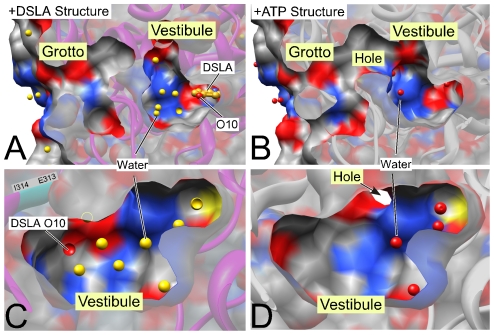
Figure 8. Ligand-induced changes in the vestibule and grotto region of luciferase.
The “active” DSLA bound (left, 2D1S.pdb) and ATP bound (right, 2D1Q.pdb) structures are compared from the same viewing points. Upper panels show a cross section through the vestibule and grotto region beyond the luciferin binding pocket (compare Fig. 7). The lower panels show the vestibule as viewed from the luciferin pocket; the left panel shows the O10 of DLSA for orientation. Water molecules are shown in red in the +ATP structure and in gold in the DSLA structure (one of the eight water molecules is partially hidden in panel C and is indicated with a circle). The cross-section surface capping is semitransparent. Surface coloring is grey for carbon, red for oxygen and blue for nitrogen. DSLA (gold carbons in A) is a surrogate for luciferin (Fig. 1B). Similar changes are seen in American luciferase (3IES.pdb and 3IEP.pdb).

