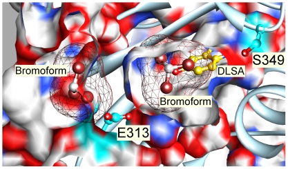
Figure 9. Relationship of photolabeled residues to other ligands bound.
The structure of American luciferase with two bromoform molecules bound (1BA3.pdb; bromine atoms are brown) was superimposed on the DSLA–bound Japanese luciferase structure (2D1S.pdb). A cross section of 2D1S is shown without surface capping in a surfaced ribbon diagram (light blue) with the photolabeled residues shown with cyan carbons. DLSA is shown with gold carbons. The surface of the bromoforms is shown in mesh representation. The carbon atom of the inner bromoform (on the right), is centered within 2 Å of the terminal oxygen (O10) of DSLA and overlaps C1, C5 and C6 of the benzothiazole ring of DSLA (see Fig. 7 for numbering). The outer bromoform (left) is situated in the grotto region 2.7 Å from Glu-313. It clashes sterically with the superimposed Japanese luciferase structure.

