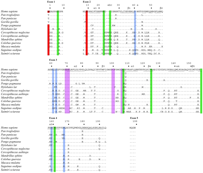
Figure 1. Alignment of primate APOBEC3A proteins.
Twelve primate sequences were compared to Homo sapiens used as reference. Only differences are shown. Hyphens denote gaps introduced to maximize sequence identity. The numbering corresponds to that of the human sequence. The letters a, b, c are added to adjacent residue to accommodate insertions. Red denotes the first (M1, exon 1) and second initiation start codons (M13, exon 2). The crucial cytidine deaminase motif residues are highlighted in magenta. Positively and negatively selected codon sites are in blue and green respectively. The predicted secondary structure motifs for hA3A are underlined.

