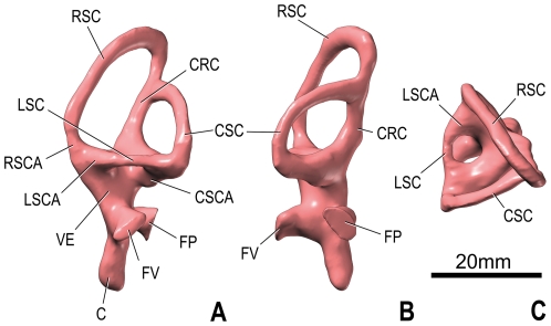
Figure 5. Endosseous labyrinth of the left inner ear of Spinophorosaurus nigerensis (GCP-CV-4229) reconstructed from CT scan; in lateral (A), caudal (B), and dorsal (D) views.
Orientations were determined based on orientation of the labyrinth within the braincase and with the lateral semicircular canal placed horizontally. Abbreviations: C, cochlea ( = lagena); CRC, crus commune; CSC, caudal ( = posterior) semicircular canal; CSCA, ampulla of caudal semicircular canal; FP, fenestra perilymphatica ( = round window); FV, fenestra vestibuli ( = oval window); LSC, lateral ( = horizontal) semicircular canal; LSCA, ampulla of lateral semicircular canal; RSC, rostral ( = anterior) semicircular canal; RSCA, ampulla of rostral semicircular canal; VE, vestibule of inner ear.

