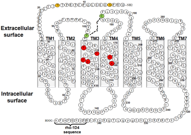
Figure 1. Two-dimensional representation of the TPα amino acid sequence.
Amino acids are shown in single-letter codes. Shown are the seven transmembrane helices, the disulphide bond between the Cys102 and Cys183 (green colored residues), the N-glycosylated residues Asn4 and Asn16 (orange colored residues), and the rho-1D4 octapeptide epitope tag at the C-terminus. The two conserved residues Ala1604.53 and Gly1644.57 on TM4 along with the residues on TM3 mutated in this study are shown in red.

