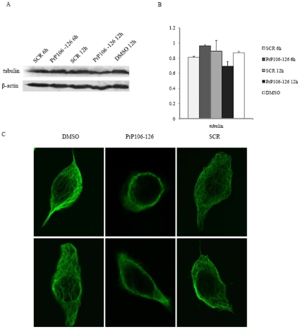
Figure 6. Analyses of the tubulin levels and microtubule structure in cells exposed to DMSO, PrP106–126 or SCR.
A. Western blots with anti-α-tubulin mAb. B. Quantitative analyses of each gray numerical value of tubulin vs that of individual β-actin. The average relative gray value is calculated from three independent blots and presented as mean ± S.D. C. Immunofluorescence images of cellular microtubule structure.

