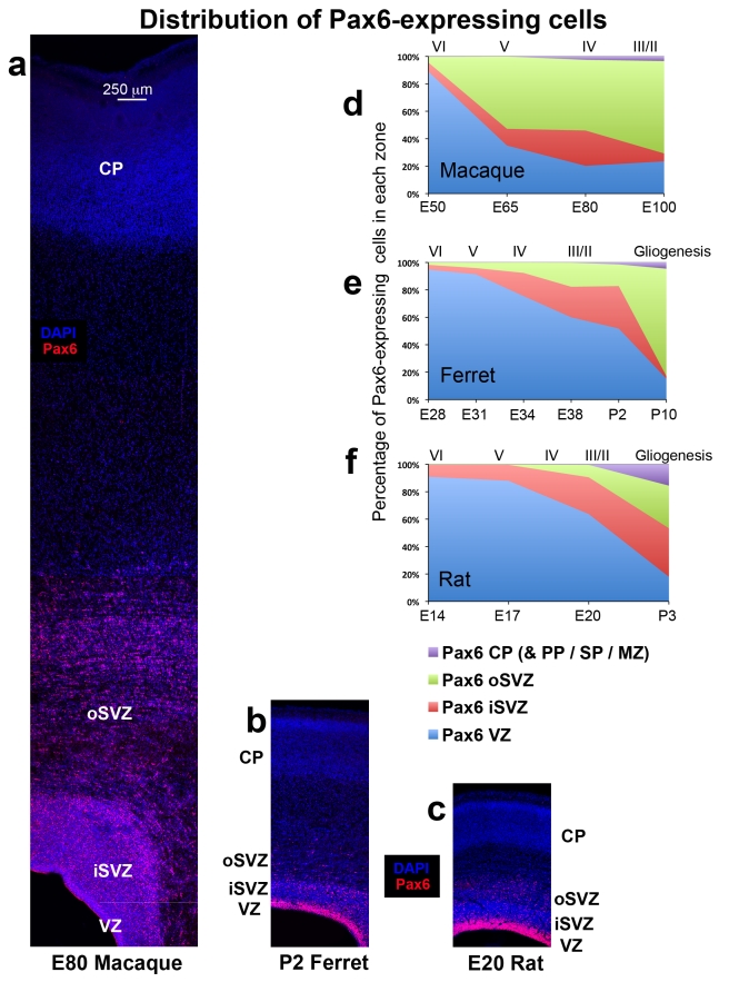
Figure 9. The distribution of Pax6+ cells during cortical development is similar in ferrets and rats, but different in macaques.
(a–c) Coronal sections of somatosensory cortex from E80 macaque, P2 ferret and E20 rat immunostained for Pax6 (red) and counterstained with DAPI (blue). Images are displayed at the same scale. In each species a dense inner band of Pax6+ cells was colocalized with the ventricular zone (VZ), and a diffuse band of Pax6-expressing cells extended outward through the inner subventricular zone (iSVZ) and the outer SVZ (oSVZ). The iSVZ and oSVZ were identified based on the pattern of DAPI staining as described above. (d–f) Graphs showing changes in the distribution of Pax6+ cells during development of the somatosensory cortex. The stage of development is shown at the bottom of the graphs, and the approximate cortical layer generated at each stage of development is indicated along the top of each graph. (d) In macaque the distribution of Pax6+ cells rapidly shifted to the oSVZ. At E50 nearly 90% of Pax6+ cells were located in the VZ. But by E65 during production of layer 5 neurons [20], the majority of Pax6+ cells (60%) were located in the oSVZ. (e) The distribution of Pax6+ cells in the gyrencephalic ferret shifted to the oSVZ much more slowly than in macaque. At P2 during genesis of layer 2 neurons [19], 52% of Pax6+ cells remained in the VZ. (f) The distribution of Pax6+ cells in the lissencephalic rat was similar to that of the gyrencephalic ferret. At the end of neurogenesis on E20 during production of layer 2 neurons [21], the majority of Pax6+ cells (64%) were still located in the VZ. Legend indicates histological zones: VZ: blue; iSVZ: red; oSVZ: green. Cortical plate (CP)/preplate (PP)/subplate (SP)/marginal zone (MZ): purple. Scale bar in (a) applies to (a–c).

