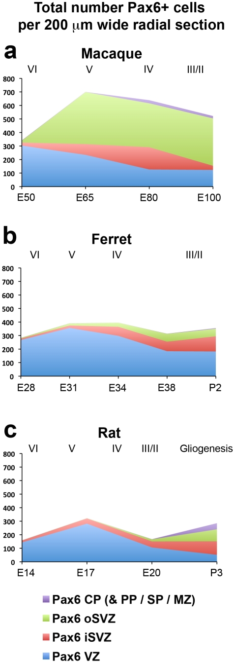
Figure 10. Graphs showing the total number of Pax6+ cells in the ventricular zone (VZ), inner subventricular zone (iSVZ), outer SVZ (oSVZ), and cortical plate (CP) within a 200 µm wide radial unit of macaque, ferret and rat somatosensory cortex.
(a–c) There was a similar number of Pax6+ cells in the iSVZ of each species, but macaque had a much larger number of Pax6+ cells in the oSVZ. The stage of development is shown at the bottom of each graph, and the approximate cortical layer generated at each stage of development is indicated along the top of each graph. Legend indicates histological zones: VZ: blue; iSVZ: red; oSVZ: green; CP: purple. PP, preplate; SP, subplate; MZ, marginal zone.

