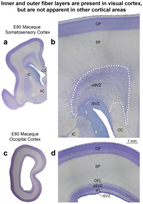
Figure 20. The inner fiber layer (IFL) and outer fiber layer (OFL) are present in the E80 macaque visual cortex but are not apparent in other cortical areas.
(a,c) Nissl-stained coronal sections of somatosensory and visual cortex taken from E80 macaque. (b,d) Higher magnification images taken from the sections shown in (a) and (c). Dotted line in (b) represents the outer boundary of the outer subventricular zone (oSVZ) in somatosensory cortex determined through Tbr2 immunostaining on adjacent sections (see Fig. 5). The outer fiber layer (OFL) and inner fiber layer (IFL) are visible in the visual cortex but are not apparent in cortical areas rostral to the occipital lobe. In somatosensory cortex the boundary between the inner SVZ (iSVZ) and oSVZ can be visualized in Nissl stained tissue as a sharp border created by differences in cell density. In the visual cortex the IFL divides the iSVZ from the oSVZ. In somatosensory cortex the oSVZ extends much farther from the ventricle than it does in visual cortex and is characterized by a striated appearance. In the E80 macaque the ventricular zone has become very thin and the cell dense proliferative zone that surrounds the lateral ventricle consists almost entirely of iSVZ. The subplate (SP) was identified according to Smart et al 2002 [9]. Scale bar in (b) applies to (d). CC, corpus callosum; IC, internal capsule; AC, anterior commissure; CP, cortical plate.

