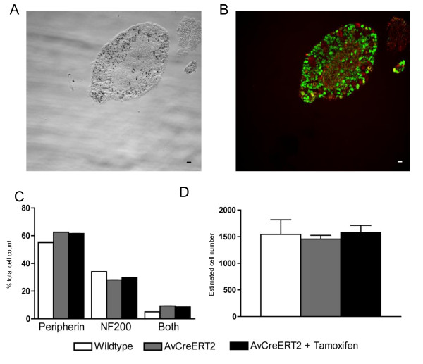
Figure 3.
Co-localization of X-gal staining with peripherin and NF200 positive neurons in the dorsal root ganglia of the adult AvCreERT2 mice. A - X-gal (transmitted light) and B. - Anti-peripherin (green) and anti-NF200 staining (red). C - Tamoxifen treatment does not alter the composition of DRG. White bars - wt, Grey bars - uninjected AvCreERT2 animals, Black bars - AvCreERT2 animals injected with tamoxifen (2 mg per day, 5 days). D - Quantification of the total number of neurons per DRG of wildtype and AvCreERT2 mice (untreated and injected with tamoxifen). Data are presented as mean ± SEM. Scale bars = 40 μm.

