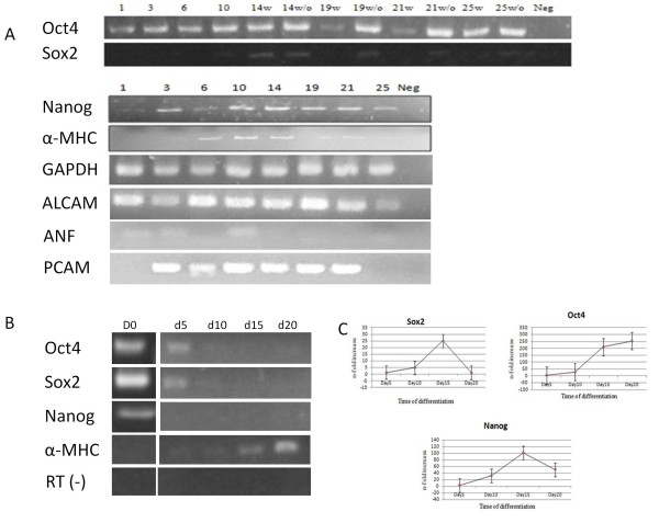
Figure 2.
Expression of pluripotency and cardiac markers during differentiation in suspension culture. A) RT-PCR showed the expression of pluripotency as well as cardiac lineage genes during differentiation in the suspension bioreactor. The G418 used from day 10 of differentiation to select cardiomyocytes: w: with drug; w/o: without drug, Neg: Negative control. The numbers show days past differentiation. Cardiac markers were examined in non-drug selected bioreactors. B) RT-PCR showed the absence of pluripotency markers gene expression during static differentiation. The expression of the cardiac lineage gene α-MHC increased during differentiation in static culture in non-drug selected cultures. C) Quantitative RT-PCR results in non-drug selected cultures revealed the fold increase of gene expression due to shear forces in suspension bioreactor compared to static culture during the time course of cardic differentiation. Values represent means±SD of 3 independent experiments. p < 0.05.

